Hidden Heparins: HIT Happens
Case Objectives
- Review the presentation of heparin-induced thrombocytopenia (HIT) and its primary complication, thrombosis.
- Discuss the management of HIT, including use of direct thrombin inhibitors and extended anticoagulation with a coumarin derivative.
- Identify safeguards that medical institutions can undertake to avoid exposure to heparin in individuals who have received the diagnosis of HIT.
Case & Commentary: Part 1
A patient with a history of end-stage renal disease requiring hemodialysis was admitted for evaluation of non-healing ulcers and leukocytosis. She had been admitted 1 month prior for evaluation of peripheral artery disease. During that hospitalization, the patient underwent angioplasty of the right femoral artery, complicated by postoperative gangrene of the right foot requiring above-the-knee amputation. She also developed axillary vein thrombosis, and ultimately a diagnosis of heparin-induced thrombocytopenia (HIT) was made. She was treated with argatroban, and it was noted on the chart that the patient should receive no further heparin.
Heparin-induced thrombocytopenia (HIT) occurs when heparin molecules stimulate formation of a pathogenic IgG antibody (HIT antibody) that leads to platelet activation (promoting thrombosis) and clearance (leading to thrombocytopenia). The typical presentation of HIT involves a hospitalized patient who develops thrombocytopenia within 5–10 days of receiving heparin; decreases in the platelet count to 9/L or by greater than or equal to 50% below the baseline value are generally considered to be significant.(1) Up to half of patients with HIT will experience thrombosis, in either the arterial or venous circulation.(3) This patient appears to have experienced thrombosis (axillary vein thrombosis, and possibly a lower extremity arterial event leading to limb ischemia and amputation) as her primary manifestation of HIT.
In a hospitalized patient, the diagnosis of HIT may be confounded by the presence of other potential causes of thrombocytopenia. Still, a pretest scoring system, such as the 4 Ts—which takes into account the degree of Thrombocytopenia, Timing of the platelet count decline in relation to heparin use, presence of any Thrombosis, and any oTher potential causes for thrombocytopenia (Figure 1)—may facilitate assignment of a diagnosis.(4) Laboratory studies such as the HIT antibody enzyme-linked immunoassay (ELISA) and the serotonin-release assay can later confirm the diagnosis, but initiation of treatment for HIT should not be delayed pending results of these specialized studies.
Unfractionated heparin (UFH) is associated with HIT more frequently than low-molecular-weight heparin (LMWH). A recent meta-analysis including 7000 medical and surgical patients who received UFH or LMWH as thromboprophylaxis showed a rate of HIT of 2.6% and 0.6%, respectively.(2) As the patient in this case had undergone angioplasty of the right femoral artery for treatment of peripheral arterial disease, it is likely that she received heparin at some point during that hospital stay.
Due to the high risk of thrombosis associated with HIT, the appropriate initial management of the condition includes discontinuation of all forms of heparin (including flushes) and initiation of an alternative anticoagulant, such as a direct thrombin inhibitor (DTI). Argatroban, lepirudin (Refludan®), and bivalirudin (Angiomax®) are DTIs that are commercially available in the United States (Table). The DTI should be continued until the platelet count has recovered and the patient is adequately anticoagulated with a coumarin derivative (warfarin). Because of the risk of venous limb gangrene that can result from transient hypercoagulability associated with early warfarin therapy in HIT patients, warfarin therapy should not be started until the platelet count recovers and then only with overlapping DTI therapy. In patients with HIT and thrombosis, as in this case, anticoagulation, usually with a coumarin derivative, should be continued for a minimum of 3–6 months following detection of the thrombosis.(5,6) Even in patients with HIT who present without thrombosis, the risk of thrombosis persists for at least 30 days, with up to 50% of patients developing venous or arterial events.(3) Therefore, patients with confirmed HIT without thrombosis should be anticoagulated (typically with a coumarin derivative) for at least 4 weeks after HIT is detected.(6)
Case & Commentary: Part 2
On this admission, the patient was found to be tachycardic and hypotensive, with excoriations of the skin over the breast, abdomen, right thigh, and gluteal region. Her labs were significant for leukocytosis of 17.1 x 10⁹/L and hypoalbuminemia of 2.4 gm/dl. The patient was started on antibiotics (amikacin and vancomycin). Blood cultures eventually grew candida, and amphotericin was added to her regimen. She appeared to be improving with a decrease in white blood cell count (WBC) to 10 x 109/L. Over the next few days, however, she developed ischemia in her right hand, which eventually became cold and pulseless. It was also noted at this time that her platelet count had dropped since admission. On hospital day 8, the leukocyte count increased again, her respiratory status worsened, and she died, presumably from overwhelming sepsis.
In light of the high risk of thrombosis that accompanies the diagnosis of HIT, it is possible that the patient’s presenting signs of tachycardia and hypotension stemmed from an HIT-related central venous or arterial thrombotic event, such as myocardial infarction or pulmonary embolism. Leukocytosis does not complicate HIT per se, but inflammation or infection from ischemia could lead to an elevated white cell count.
Shortly after admission, this patient did develop signs of arterial insufficiency with right hand ischemia that progressed to pulselessness. Although HIT is complicated by symptomatic venous thrombosis more often than arterial thrombosis (7), arterial thrombosis may occur more frequently in certain patients with HIT, such as individuals with cardiovascular disease.(8)
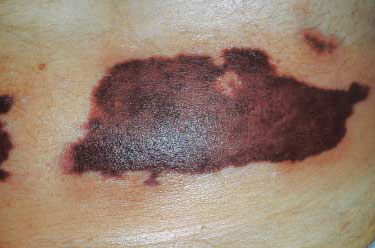
Interestingly, this patient presented with “excoriations” of the skin. Up to 10%–20% of patients receiving subcutaneous UFH or LMWH who develop HIT will first have erythematous, nodular, plaque-like, or necrotic lesions (Figure 2) at the injection site.(7) Although HIT antibodies are detectable in most of these patients, thrombocytopenia is present in only a minority.(9) We are not told that heparin was ever injected subcutaneously in this case, and the location of some of the lesions (e.g., breast) makes an injection-related phenomenon less likely, although skin lesions that occur at sites that are distant from the location of subcutaneous injection have been reported.(10) Dermal necrosis also has been reported following use of intravenous heparin.(11)
In contrast, coumarin-induced skin necrosis (CISN) rarely occurs when warfarin is introduced in a patient with HIT and typically begins within days of starting the oral anticoagulant. A proposed mechanism for the condition involves worsening of HIT-induced hypercoagulability due to an acute reduction in levels of protein C or S. Localized, progressive, microvascular thrombosis ensues that typically leads to necrosis of the dermis overlying the breast, abdomen, thigh, or leg.(7) We are not told that this patient ever received warfarin, but the dermatologic “excoriation” may well have been a skin manifestation of HIT.
Case & Commentary: Part 3
Autopsy revealed thrombi in the vessels of the skin of breast and abdomen. A thorough review of the chart and hemodialysis records revealed that, during this second hospitalization, the patient had been repeatedly exposed to heparin during dialysis sessions, despite her recent history of HIT and the chart notes to avoid heparins.
The histologic finding at autopsy of microthrombi in dermal vessels strongly suggests that the skin lesions that were initially observed over the breast, abdomen, and thigh represented sequelae of ongoing HIT, which probably had developed following exposure to heparin during the first admission to the hospital. Although HIT had been documented and treated during that hospitalization, the patient continued to receive heparin in the hemodialysis suite.
A diagnosis of HIT does not necessarily obviate any future use of heparin, but several factors must be carefully considered before heparin is given. Although certain patients may experience no recurrence of thrombocytopenia upon re-challenge with heparin as soon as 1.5 months after an episode of HIT (1), most patients with HIT continue to test positive for HIT antibodies well after the thrombocytopenia has resolved. When measured by ELISA, the median duration of HIT antibody positivity is 85 days.(1) This observation has led to the usual practice of waiting at least 100 days before considering use of heparin in patients with a prior history of HIT and documenting negativity for the HIT antibody before re-exposing to heparin. If re-exposure is absolutely necessary (i.e., for vascular surgery), short-term use of the heparin may be considered with frequent monitoring of the platelet count. Patients with persistent HIT antibodies, as our patient most likely had, who are re-challenged with heparin can develop rapid-onset HIT, in which thrombocytopenia and thrombosis develop with alarming speed, sometimes as soon as a day after re-exposure to heparin.(7)
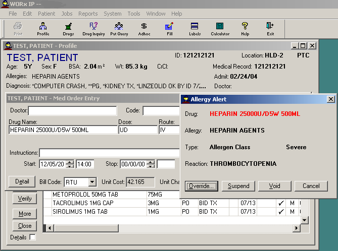
This case underscores the need for safeguards for patients who are diagnosed with HIT to prevent “accidental” exposure to heparin. In our hospital (UCSF Medical Center), the electronic pharmacy profile (WORx, Mediware Information Systems, Lexena, Kansas) of any patient in whom HIT is diagnosed is updated to indicate the diagnosis. If a heparin (UFH or LMWH) subsequently is ordered by a care provider, an alert is generated (Figure 3) that is visible to the hospital pharmacist, who contacts the ordering provider to advise of the prior history. In this regard, patients with a prior history of HIT are “protected” from orders for UFH or LMWH that are written without a knowledge of the patient’s history of HIT, and the protection extends to future admissions. As an additional precaution, a placard indicating “Heparin-Induced Thrombocytopenia: No Heparin” is placed above the bedside in rooms of inpatients who receive the diagnosis, and an allergy is recorded in the medical record.
Unfortunately, in most hospitals, heparins (typically UFH) are stocked in more locations than simply the clinical pharmacy, and patients can be exposed to the drug, usually as part of a “routine” maneuver such as flushing a venous access device. Administration of heparin in quantities as small as those routinely used to flush central venous catheters and other vascular access devices can lead to HIT antibody formation or HIT itself (12–14), and minor exposure via this route in a patient with persistently circulating HIT antibodies can promote re-emergence or worsening of thrombocytopenia or thrombosis.(15) To reduce the risk of promoting or exacerbating HIT through use of heparin flushes of invasive catheters, some institutions have adopted a policy of routinely using normal saline for their flushes. Such an approach is supported by several studies, including a randomized controlled trial of UFH versus saline flushes for invasive catheters used in the operating suite and a meta-analysis of patency rates of peripheral venous catheters flushed with UFH or normal saline, which found no benefit to UFH for this purpose.(12,16) Heparin is still used routinely to prevent thrombosis of the hemodialysis circuit, however. Because heparin usually is stocked in the dialysis unit, the pharmacy and aforementioned system of alerts can be bypassed.
The significant risk of HIT and its devastating sequelae has prompted consideration of policies that aim to remove heparin from all patient care areas. When such policies are implemented, ordering heparin requires an MD order to the pharmacy, even when the heparin is for flushing of catheters. Implementation of computerized electronic alerts, especially if linked to physician ordering screens, may provide the best method of risk notification, as has been demonstrated in other applications.(18)
Take-Home Points
- HIT can occur in medical or surgical patients after exposure to UFH or LMWH in any form (subcutaneous injection, intravenous, flushes).
- The clinical presentation of HIT can include isolated thrombocytopenia, isolated arterial or venous thrombosis, or both conditions. Skin lesions occur in a minority of patients with HIT but increase the probability of the diagnosis in a thrombocytopenic patient who has been exposed to heparin.
- Patients with a history of HIT should generally not be re-exposed to heparin (either UFH or LMWH), but when clinically indicated, heparin may be used for a short period of time provided HIT antibodies are no longer detectable.
- All patients with a prior history of HIT should have the medical and pharmacy record permanently amended to indicate the diagnosis.
- Identifying uses of heparins that circumvent provider orders can lower the risk that a patient with HIT (or at risk for HIT) will be exposed to heparin.
Patrick F. Fogarty, MD Director, Hemostasis and Thrombosis Clinic Department of Medicine, Division of Hematology/Oncology University of California, San Francisco
Faculty Disclosure: Dr. Fogarty has declared that neither he, nor any immediate member of his family, has a financial arrangement or other relationship with the manufacturers of any commercial products discussed in this continuing medical education activity. The commentary does include information regarding off-label use of bivalirudin for treatment of HIT. All conflicts of interest have been resolved in accordance with the ACCME Updated Standards for Commercial Support.
References
1. Warkentin TE, Kelton JG. Temporal aspects of heparin-induced thrombocytopenia. N Eng J Med. 2001;344:1286–1292. [go to PubMed]
2. Martel N, Lee J, Wells PS. Risk for heparin-induced thrombocytopenia with unfractionated and low-molecular-weight heparin thromboprophylaxis: a meta-analysis. Blood. 2005;106:2710–2715. [go to PubMed]
3. Warkentin TE, Kelton JG. A 14-year study of heparin-induced thrombocytopenia. Am J Med. 1996;101:502–507. [go to PubMed]
4. Lo GK, Juhl D, Warkentin TE, Sigouin CS, Eichler P, Greinacher A. Evaluation of pretest clinical score (4 T’s) for the diagnosis of heparin-induced thrombocytopenia in two clinical settings. J Thromb Haemost. 2006;4:759–765. [go to PubMed]
5. Fogarty PF, Dunbar CE. Thrombocytopenia. In: Rodgers GP, Young NS, eds. Bethesda Handbook of Hematology. Philadelphia, PA: Lippincott, Williams and Wilkins; 2005:256–257.
6. Alving BM. How I treat heparin-induced thrombocytopenia and thrombosis. Blood. 2003;101:31–37. [go to PubMed]
7. Warkentin TE. Clinical picture of heparin-induced thrombocytopenia. In: Warkentin TE, Greinacher A, eds. Heparin-Induced Thrombocytopenia. 3rd ed New York, NY: Marcel Dekker; 2004:53-106.
8. Boshkov LK, Warkentin TE, Hayward CP, Andrew M, Kelton JG. Heparin-induced thrombocytopenia and thrombosis: clinical and laboratory studies. Br J Haematol. 1993;84:322–328. [go to PubMed]
9. Warkentin TE, Roberts RS, Hirsh J, Kelton JG. Heparin-induced skin lesions and other unusual sequelae of the heparin-induced thrombocytopenia syndrome: a nested cohort study. Chest 2005;127:1857–1861. [go to PubMed]
10. Balestra B, Quadri P, Dermarmels Biasiutti F, Furlan M, Lämmle B. Low molecular weight heparin-induced thrombocytopenia and skin necrosis distant from injection sites. Eur J Haematol. 1994;53:61–63. [go to PubMed]
11. Hartman AR, Hood RM, Anagnostopoulos CE. Phenomenon of heparin-induced thrombocytopenia associated with skin necrosis. J Vasc Surg. 1988;7:781–784. [go to PubMed]
12. Warkentin TE, Ling E, Ho A, Sheppard JI. “Incidental” unfractionated heparin (UFH) vs normal saline (NS) flushes for intraoperative invasive catheters and the frequency of formation of heparin-induced thrombocytopenia IgG antibodies (HIT-IgG): a randomized, controlled trial. Blood. 1998;92(suppl 1):91b.
13. Brushwood DB. Hospital liable for allergic reaction to heparin used in injection flush. Am J Hosp Pharm. 1992;49:1491–1492. [go to PubMed]
14. Heeger PS, Backstrom JY. Heparin flushes and thrombocytopenia. Ann Intern Med. 1986;105:143. [go to PubMed]
15. Rice L, Jackson D. Can heparin cause clotting? Heart Lung. 1981;10:331–335. [go to PubMed]
16. Randolph AG, Cook DJ, Gonzalez CA, Andrew M. Benefit of heparin in peripheral venous and arterial catheters: systematic review and meta-analysis of randomised controlled trials. BMJ. 1998;316:969–975. [go to PubMed]
17. Vanholder RC, Camez AA, Veys MM, et al. Recombinant hirudin: a specific thrombin inhibiting anticoagulant for hemodialysis. Kidney Int. 1994;45:1754–1759. [go to PubMed]
18. Kucher N, Koo S, Quiroz R, et al. Electronic alerts to prevent venous thromboembolism among hospitalized patients. N Engl J Med. 2005;352:969–977. [go to PubMed]
Table
Direct thrombin inhibitors.
| Agent | Description | Indication | Dosing | Comment | ||||
|---|---|---|---|---|---|---|---|---|
| Argatroban | Synthetic direct thrombin inhibitor | Prophylaxis or treatment of HIT, including post–percutaneous coronary intervention | Obtain baseline PTT. Start continuous infusion at 2 m/kg/min. Titrate to achieve PTT of 1.5–3 times the baseline value. Do not allow PTT to exceed 100 seconds, nor the infusion rate to exceed 10 mg/kg/min. |
|
||||
| Lepirudin (Refludan®) | Recombinant hirudin; direct thrombin inhibitor | Treatment of HIT with associated thrombosis | Obtain baseline PTT. Give slow bolus of 0.4 mg/kg then continuous infusion of 0.15 mg/kg/hr. Titrate to achieve PTT of 1.5–2.5 times baseline value. PTTs should be obtained 4 hours after starting the infusion and at least daily during treatment. |
|
||||
| Bivalirudin (Angiomax®) | Semi-synthetic derivative of hirudin; direct thrombin inhibitor | Unstable angina in patients undergoing percutaneous transluminal coronary angioplasty | 1.0 mg/kg IV bolus followed by 2.5 mg/kg/hr infusion for 4 hours; may continue infusion at 0.2 mg/kg/hr for up to 20 hours. |
|
Figures
Figure 1. A Clinical Pretest Approach to the Diagnosis of HIT (Adapted from Lo et al.[4]).
| Characteristic of Heparin-Induced Thrombocytopenia (HIT) | Score (Fill in 0–2) | Pretest Probability Score | ||
| 2 | 1 | 0 | ||
| Thrombocytopenia (x 109/L) | ____ | Nadir 20–100, or >50% platelet fall from baseline | Nadir 10–19, or 30–50% platelet fall from baseline | Nadir |
| Timing of onset of platelet fall after starting heparin | ____ | Day 5–10, or less than or equal to day 1 if recent heparin* | >Day 10, or timing uncertain but compatible with HIT | Less than or equal to Day 1 (no recent heparin) |
| Thrombosis/additional manifestations | ____ | Proven thrombosis, skin necrosis, or ASR† | Progression or recurrence of established thrombosis; silent thrombosis; erythematous skin lesions | None |
| Other potential causes of platelet decline | ____ | None evident | Possible | Definite |
| Total Pretest Probability Score | ____ | See “Total PreTest Probability Score,” below |
| Total Pretest Probability Score | ||||||||
| High | Moderate | Low | ||||||
| 8 | 7 | 6 | 5 | 4 | 3 | 2 | 1 | 0 |
| Stop heparin‡, give alternative non-heparin anticoagulant Argatroban or lepirudin (or bivalirudin) Avoid warfarin** | (Individualize management) | Continue (LMW) heparin |
*Recent heparin = exposure within the past 30 days (2 points) or past 30–100 days (1 point). †ASR = acute systemic reaction, including hypertension, tachypnea, fever, following intravenous heparin bolus. ‡Discontinue all forms of heparin, including vascular catheter heparin flushes and heparin-coated catheters. **Warfarin should not be started until platelet count has recovered (plt >100K); starting dose should be low, and therapy should be overlapped with a direct thrombin inhibitor for a minimum of 5 days.
Figure 2. Heparin-Induced Skin Necrosis. (Picture reprinted with permission from International Journal of Dermatology.)
Figure 3. Example of an Electronic Alert upon Attempting to Order Heparin in a Patient with a Heparin Allergy: WORx® Software.


