Overdose on Oxygen?
The Case
An 84-year-old woman with a history of chronic obstructive pulmonary disease (COPD) on home oxygen (1–2 L/min) presented to the emergency department with cough, chills, and shortness of breath. On physical examination, she was afebrile and tachypneic, and her oxygen saturation was 94% on 2 L of oxygen. Her pulmonary examination was unremarkable. The chest radiograph revealed bilateral lower lobe infiltrates. The patient was admitted with a diagnosis of community-acquired pneumonia and a COPD exacerbation. She was given appropriate therapies and, with her mild illness, was expected to be discharged the following day.
On the morning following admission, she was found to be somnolent and minimally arousable. She remained afebrile with a normal blood pressure and pulse, and the remainder of the physical examination was unchanged. The physician on duty noted that her oxygen saturation was 98% and she was receiving oxygen by nasal cannula at a rate of 4 L/min. An arterial blood gas was performed, which showed a pH of 7.21 (normal, 7.35–7.45) with a partial pressure of CO2 (PaCO2) of 101 mm Hg (normal, 35–45 mm Hg) and an oxygen of 85 mm Hg. The hypercarbia (increased CO2) was felt to be due to excessive oxygen administration and likely explained her change in mental status.
She was immediately transferred to the intensive care unit (ICU) where she failed noninvasive positive pressure ventilation (NIPPV) and required endotracheal intubation. Her oxygen "dose" was lowered, in an effort to keep her saturation in the 90%–94% range. With that (and treatment of her infection and COPD), her hypercarbia promptly resolved. She was extubated the following day, had an uncomplicated subsequent hospital course, and was discharged home 3 days later.
The Commentary
This case raises concerns about the assessment, management, and monitoring of a patient with one of the most common medical conditions seen in hospital emergency departments—exacerbation of COPD. In particular, this case highlights the challenge of appropriate oxygen delivery in the setting of a COPD exacerbation.
The admitting physicians were aware that the patient was on chronic home oxygen therapy for her COPD. As home oxygen is generally only prescribed for patients whose blood oxygen tension when breathing air is below 55 mm Hg, the physicians could assume that her baseline oxygen saturation was 88% or less without oxygen (Table).(1) Although her baseline oxygen saturation while on 2 L/min was likely not known by the admitting team, they may have been falsely reassured by the oxygen saturation of 94%. Unfortunately, a blood gas was not done in the emergency department, despite the fact that most expert guidelines recommend blood gases on all patients with exacerbations of COPD.(2) A blood gas would have provided valuable information about her blood pH as well as levels of CO2, possibly identifying her as a patient at risk for hypercarbia (elevated levels of CO2).
The currently recommended target oxygen tension in exacerbated COPD is about 60–65 mm Hg, which is equivalent to a saturation of approximately 90%–92% (Table).(2) Despite an initial blood oxygen saturation of 94%, this patient's oxygen flow rate was increased from 2 to 4 L/min. This is equivalent to increasing the FIO2 from approximately 28%–35%.(3) The increased dose of oxygen raised her oxygen saturation from 94%–98%, significantly above published guidelines and equivalent to increasing the blood oxygen tension from about 70 mm Hg to about 100 mm Hg (Table). This increased oxygen tension likely led to progressive hypercapnia (increasing CO2 level) overnight (see below for further explanation of the risks of excess oxygen in patients with severe COPD). Unfortunately, there appears to have been no further assessment of her condition until she was found to be semicomatose with a high CO2 and a respiratory acidosis on the morning following admission.
Why do we care about hypercapnia and respiratory acidosis? Respiratory acidosis has been shown to be associated with an increased risk of intubation and death in acute exacerbations of COPD.(4,5) It is possible that a blood gas at the time of admission might have shown a pH in the region of 7.30–7.35, which might have prompted the clinicians to initiate a trial of NIPPV. By the next morning, her extremely low pH of 7.21 mandated invasive ventilation, adding greatly to the risks and costs of managing her case. Her condition stabilized rapidly with controlled oxygen therapy.
As an aside, the case history does not state if the increased oxygen dose was prescribed by the admitting team or left to the discretion of the nursing staff. Audits in several countries have shown that oxygen is poorly prescribed and poorly administered in hospital settings.(6,7) There are no existing US or international guidelines for emergency oxygen therapy, but the UK guideline, which will cover all aspects of emergency oxygen use, will be published in mid-2008.(7)
Titrating oxygen appropriately in exacerbations of COPD is challenging as patients in this setting can be harmed by too much or too little oxygen. It must be remembered that most such patients (as in the present case) spend much of their lives with an abnormally low blood oxygen tension and have adjusted physiologically; their bodies become accustomed to lower oxygen levels than patients without COPD.(1,4) A review of the literature suggests that most patients with exacerbated COPD are adequately oxygenated if the oxygen tension can be maintained above 50 mm Hg, corresponding to an oxygen saturation above about 85% (contrast this to people without COPD, who require oxygen levels above 60 mm Hg or 90%).(4) The new British Thoracic Society Emergency Oxygen guideline for the United Kingdom will recommend that the saturation should be maintained above 88% in most cases of exacerbated COPD to allow an additional margin of safety.(7) However, high-risk patients with prior hypercapnic respiratory failure may be safely managed with an oxygen saturation in the range of 85%–88%.
What are the harms of excessive oxygen administration? High doses of oxygen can cause a number of adverse effects including absorption atelectasis and increased mismatch between ventilation and perfusion within the lung, which impairs elimination of carbon dioxide and thus leads to acidosis.(4,8) Whereas it was believed for many years that the "suppression of hypoxic drive" (loss of the normal increase in respiratory drive in the setting of hypoxia) was the most important cause of an increased carbon dioxide level in patients with exacerbated COPD, most, but not all, recent authors believe that other factors such as mismatched ventilation and perfusion are more important.(7) What is beyond dispute is that some (but not all) patients with exacerbated COPD will develop hypercapnia (increased CO2) if given high levels of supplemental oxygen. A study of 972 patients with this condition showed that almost half were hypercapnic on arrival in the emergency department, one-fifth were acidotic (pH below 7.35), and 4.6% were severely acidotic with pH below 7.25. The patients with the highest blood oxygen tensions had the worst acidosis, supporting the premise that excessive oxygen can lead to respiratory acidosis in this setting.(5) More than half of the patients with PaO2 above 75 mm Hg were acidotic. Based on their findings, the authors recommended keeping the oxygen saturation below 92% to avoid this risk. In a more recent study, Joosten and colleagues demonstrated that an oxygen tension above 74.5 mm Hg in acute COPD was associated with an increased risk of requirement of NIPPV and greater length of hospital stay.(9)
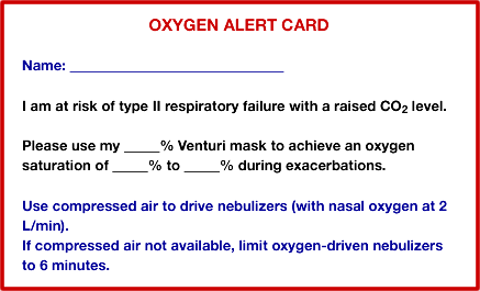
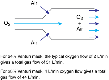
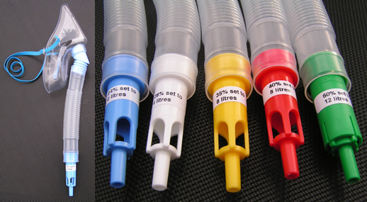
Based on these studies and others, the UK Emergency Oxygen Guideline will recommend a target oxygen saturation range of 88%–92% for most patients with exacerbated COPD.(7) Selected patients with a history of respiratory acidosis may require a lower target range such as 85%–90% based on the results of previous blood gas analysis. These patients will be issued an "oxygen alert card" (Figure 1) with individualized instructions for oxygen therapy during future exacerbations. The card should be carried by the patients at all times and made available to health care providers to help guide oxygen therapy. It is also helpful to issue such patients with a personal 24% or 28% Venturi mask for use during exacerbations.(10) Most simple oxygen masks deliver an increased dose of oxygen if the flow rate is increased, and this can be dangerous for vulnerable patients with COPD. However, a Venturi mask uses a special valve that entrains air using the Venturi principle (Figure 2). This delivers a fixed dose of oxygen even if the flow rate is increased and thus helps to ensure that the patient receives a constant and safe dose of oxygen.
The UK Emergency Oxygen Guideline will recommend that the initial treatment for patients with exacerbated COPD should be a 28% Venturi mask at 4 L/min but a 24% Venturi mask should be used if the saturation rises above 92%.(7) Blood gases should be checked in all such cases on arrival in the emergency department, and NIPPV should be instituted if the patient is acidotic (pH below 7.35).
Managing oxygen appropriately in the setting of a COPD exacerbation can be challenging. Obviously, for the hypoxic patient, oxygen can be life-saving. But clinicians must be aware of the challenges and risks of oxygen therapy in patients with severe COPD and take advantage of published guidelines to help ensure the safest and most appropriate care.
Take-Home Points
- Appropriate and safe management of oxygen therapy in the setting of COPD exacerbations is challenging.
- Oxygen tensions above about 50 mm Hg (saturation above about 85%) will protect patients from hypoxic injury during exacerbations of COPD.
- Oxygen tensions above about 75 mm Hg (saturation above about 95%) are associated with increased risk of hypercapnia and acidosis in exacerbated COPD.
- Oxygen is best prescribed to achieve a desirable target range rather than a fixed dose of oxygen. For most COPD patients, a target saturation range of 88%–92% will avoid the risks of hypoxia and hypercapnia.
- Some patients with previous episodes of respiratory acidosis may require an "oxygen alert card" with a lower (personalized) target saturation range.
B. Ronan O'Driscoll, MD Consultant Respiratory Physician Salford Royal University Hospital Salford, United Kingdom
References
1. Cranston JM, Crockett AJ, Moss JR, Alpers JH. Domiciliary oxygen for chronic obstructive pulmonary disease. Cochrane Database Syst Rev. 2005:CD001744. [go to PubMed]
2. Stoller JK. Acute exacerbations of chronic obstructive pulmonary disease. N Engl J Med. 2002;346:988-994. [go to PubMed]
3. Waldau T, Larsen VH, Bonde J. Evaluation of five oxygen delivery devices in spontaneously breathing subjects by oxygraphy. Anaesthesia. 1998;53:256-263. [go to PubMed]
4. Murphy R, Driscoll P, O'Driscoll R. Emergency oxygen therapy for the COPD patient. Emerg Med J. 2001;18:333-339. [go to PubMed]
5. Plant PK, Owen JL, Elliott MW. One year period prevalence study of respiratory acidosis in acute exacerbations of COPD: implications for the provision of non-invasive ventilation and oxygen administration. Thorax. 2000;55:550-554. [go to PubMed]
6. Boyle M, Wong J. Prescribing oxygen therapy. An audit of oxygen prescribing practices on medical wards at North Shore Hospital, Auckland, New Zealand. N Z Med J. 2006;119:U2080. [go to PubMed]
7. O'Driscoll BR, Howard L, Davison AM. British Thoracic Society. Guideline for emergency oxygen use in adult patients. Thorax. 2008:63:849-850. [go to PubMed]
8. Downs JB. Has oxygen administration delayed appropriate respiratory care? Fallacies regarding oxygen therapy. Respir Care. 2003;48:611-620. [go to PubMed]
9. Joosten SA, Koh MS, Bu X, Smallwood D, Irving LB. The effects of oxygen therapy in patients presenting to an emergency department with exacerbation of chronic obstructive pulmonary disease. Med J Aust. 2007;186:235-238. [go to PubMed]
10. Gooptu B, Ward L, Ansari SO, Eraut CD, Law D, Davison AG. Oxygen alert cards and controlled oxygen: preventing emergency admissions at risk of hypercapnic acidosis receiving high inspired oxygen concentrations in ambulances and A&E departments. Emerg Med J. 2006;23:636-638. [go to PubMed]
11. Severinghaus JW. Simple, accurate equations for human blood O2 dissociation computations. J Appl Physiol. 1979;46:599-602. [go to PubMed]
Table
Table. Approximate Relationship Between Arterial Hemoglobin Oxygen Saturation and Arterial Oxygen Tension in mm Hg at Normal pH
| Oxygen tension (mm Hg) | 50 | 55 | 60 | 65 | 70 | 75 | 80 | 85 | 90 | 95 | 100 | 105 | 110 |
|---|---|---|---|---|---|---|---|---|---|---|---|---|---|
| Oxygen saturation (%) | 85.1 | 88.3 | 90.7 | 92.4 | 93.8 | 94.9 | 95.7 | 96.6 | 97.0 | 97.5 | 97.9 | 98.2 | 98.4 |
Derived from equations in (11).
Figures
Figure 1. Example of Oxygen Alert Card. (Courtesy of Dr. O'Driscoll.)
Figure 2. Venturi Mask: Principles and Photographs. (Courtesy of Dr. O'Driscoll.)


